
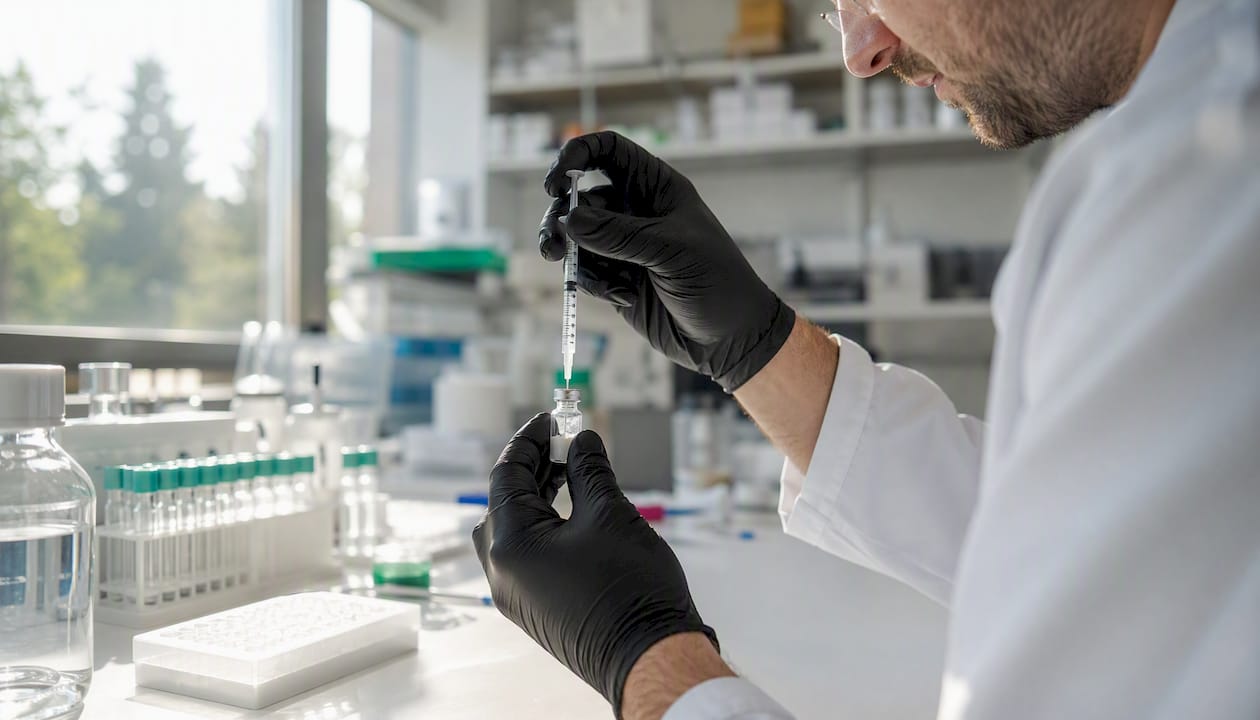
To reconstitute a lyophilised peptide correctly, draw the calculated volume of sterile solvent into a syringe and inject it slowly down the inner glass wall of the vial at an angle, directing the liquid away from the powder. Do not allow the jet to strike the cake directly, as this causes foaming and mechanical shear that can degrade the sequence. Once the solvent is added, swirl the vial gently and allow it to stand at room temperature until the solution clears. Do not vortex. Label the vial immediately with the compound name, concentration in mcg/mL, volume added, date, and your initials, then refrigerate at 2–8 °C. For multi-dose research vials, bacteriostatic water (0.9% benzyl alcohol) is the standard solvent; poorly soluble or hydrophobic peptides may require initial dissolution in HPLC-grade DMSO before aqueous dilution, and basic, aggregation-prone sequences often respond better to dilute acetic acid or HCl. All peptides supplied by Veridianlabs carry a Certificate of Analysis (COA) confirming ≥98% HPLC purity and independent mass-spectrometric identity verification; record the COA lot number in your laboratory notebook or LIMS entry at the point of reconstitution.
Research-grade reconstitution begins before the syringe is drawn. Verifying net peptide content, HPLC purity, and solvent compatibility from the COA is the first procedural step, not an administrative afterthought. A vial reconstituted without that information cannot be dosed with confidence.
This article provides general scientific information for qualified researchers. It does not constitute professional regulatory or safety advice; researchers should confirm current COSHH requirements and institutional governance obligations with their institutional safety officer and the relevant primary sources.
Table of Contents
- What materials do you need before you start?
- Which solvent should you use for reconstitution?
- How do you calculate concentration and convert to insulin syringe units?
- How do you handle poorly soluble peptides and exchange DMSO into aqueous buffers?
- What are the recommended storage conditions after reconstitution?
- What should you do when reconstitution goes wrong?
- What UK regulatory and safety obligations apply to research peptides?
- How do you read a Certificate of Analysis and evaluate a supplier?
- Key takeaways
- Why reconstitution quality depends on what is in the vial before you start
- Veridianlabs: research-grade peptides with independent batch verification
- Useful sources and further reading
What materials do you need before you start?
Assembling the correct consumables before touching the vial prevents the most common preparation errors. The table below lists every item, its specification, and the reason it matters.
| Item | Specification | Purpose |
|---|---|---|
| Peptide vial | As supplied, lyophilised | Source compound |
| Bacteriostatic water (BAC) | 0.9% benzyl alcohol, sterile | Multi-dose reconstitution solvent |
| Sterile water for injection | Single-use ampoule | Single-use reconstitution only |
| HPLC-grade DMSO | ≥98% purity | Initial dissolution of hydrophobic sequences |
| Dilute acetic acid or HCl | 0.1–1% v/v aqueous | Solubilisation of basic/aggregation-prone peptides |
| Insulin syringes | U-100 (100 units/mL) | Accurate volume measurement and dosing |
| Drawing syringe and needle | 1–3 mL, 22 G | Transferring solvent to vial |
| Alcohol swabs | 70% isopropanol | Stopper sanitisation |
| Sharps bin | UN-approved, labelled | Safe disposal of used needles |
| Fine permanent marker and labels | Waterproof | Vial identification |
| Refrigerator | Calibrated to 2–8 °C | Storage after reconstitution |
| Laminar flow bench or clean area | Per institutional SOP | Aseptic technique |
| COA and lab notebook or LIMS | Current batch documentation | Traceability and concentration calculation |
The COA is not optional documentation to file after the fact. Net peptide content and declared mass from the COA are the inputs to the concentration calculation; without them, any mcg/mL figure is an estimate. Where institutional SOPs require a laminar flow bench, that requirement takes precedence over convenience.
Pro Tip: Calibrate or verify your syringe against a known volume before the first use of a new batch. A 1% volume error in a 1 mL draw translates directly into a 1% concentration error that compounds across every subsequent dose.
Which solvent should you use for reconstitution?
Solvent selection is driven by the peptide’s net charge, hydrophobicity, and the chemistry of the downstream assay. Published methodology literature frames this decision around three physicochemical properties: solubility in aqueous media, tendency to aggregate, and sensitivity to acid-catalysed cleavage. The table below summarises the four main options and their appropriate contexts.
| Solvent | Composition | When to use | Key constraints |
|---|---|---|---|
| Bacteriostatic water (BAC) | Sterile water + 0.9% benzyl alcohol | Multi-dose vials; most hydrophilic sequences | Not for single-use-only protocols; benzyl alcohol may affect some assays |
| Sterile water for injection | Preservative-free | Single-use, immediate application | No antimicrobial protection; discard after opening |
| HPLC-grade DMSO | HPLC-grade dimethyl sulphoxide | Hydrophobic sequences that will not dissolve in aqueous media | High CD background; promotes Met/Cys oxidation; limit final % in cell assays |
| Dilute acetic acid or HCl | 0.1–1% v/v aqueous | Basic, aggregation-prone sequences | Avoid prolonged storage; acid-catalysed cleavage risk increases with time |
Bacteriostatic water is the standard choice for multi-dose research vials because its 0.9% benzyl alcohol preservative inhibits microbial growth over the opened vial’s in-use period. Sterile water, which lacks any preservative, should be used only when the entire reconstituted volume will be consumed in a single session.
DMSO requires particular care. It is an effective co-solvent for hydrophobic sequences, but it produces high background absorbance in circular dichroism (CD) spectroscopy and can promote oxidation of methionine and cysteine residues. The standard practice is to dissolve the peptide in the minimum volume of DMSO needed to achieve clarity, then dilute into aqueous buffer. For most cell-based assays, the final DMSO concentration should remain at or below 0.1–0.5% v/v; exceeding this range risks cytotoxic artefacts that confound results. Dilute acetic acid (typically 0.1% v/v) works well for basic sequences but should not be used as a long-term storage vehicle, as prolonged acid exposure increases the risk of acid-catalysed peptide bond cleavage.
Pro Tip: When in doubt about solubility, add a small volume of DMSO first to wet the powder, then dilute with aqueous buffer. This two-step approach resolves most solubility problems without requiring a full DMSO stock.

How do you calculate concentration and convert to insulin syringe units?
The concentration calculation follows a single formula: convert the vial’s net peptide content from milligrams to micrograms, divide by the solvent volume in millilitres to obtain mcg/mL, then convert mL to insulin units using the relationship 100 units = 1.0 mL.
Formula:
- Net peptide content (mg) × 1000 = total mcg
- Total mcg ÷ solvent volume (mL) = concentration (mcg/mL)
- Desired dose (mcg) ÷ concentration (mcg/mL) = volume (mL)
- Volume (mL) × 100 = units on a U-100 insulin syringe
The worked examples below cover three common vial configurations.
| Vial content | Solvent added | Concentration | dose varies depending on application |
500 mcg dose |
| — | — | — | — | — |
| 5 mg | 2 mL | 2,500 mcg/mL | 10 units | 20 units |
| 5 mg | 5 mL | — | 25 units | 50 units |
| 10 mg | 2 mL | 2,500 mcg/mL | 5 units | 10 units |
Choosing a solvent volume that places typical doses in the 10–50 unit range on a U-100 syringe reduces pipetting error and improves reproducibility across users. A dose of 5 units or fewer is difficult to measure accurately with a standard insulin syringe; a dose above 50 units occupies a large fraction of the syringe barrel and leaves little room for error correction. The 5 mg into 2 mL configuration (2,500 mcg/mL) is a practical starting point for many research sequences, but the final volume must always be calculated from the COA’s net peptide content rather than the nominal labelled mass. Purity below 98% and moisture content both reduce the actual peptide mass available, and using the nominal figure without adjustment will overestimate concentration.
For vial concentration standardisation and reproducibility considerations, particularly when comparing results across laboratories or experimental runs, documenting the exact net peptide content used in each calculation is a minimum requirement for auditable research records.
How do you handle poorly soluble peptides and exchange DMSO into aqueous buffers?
Hydrophobic sequences that resist aqueous dissolution require a two-stage approach. The standard procedure is to dissolve the peptide in the minimum volume of HPLC-grade DMSO needed to achieve a clear solution, then dilute stepwise into the target aqueous buffer while monitoring for precipitation. Keeping the final DMSO concentration at or below 0.1–0.5% v/v is the accepted limit for most cell-based assays, though the specific tolerance depends on the cell line and assay format.
For CD spectroscopy, DMSO presents a more fundamental problem. Peptides dissolved in DMSO yield high background absorbance in CD spectra, masking the characteristic helical or sheet signatures that the technique is designed to detect. A published solvent-exchange protocol addresses this using a vapour-diffusion method with ammonium nitrate (NH4NO3).
The NH4NO3 vapour-diffusion method involves placing a concentrated NH4NO3 solution (60% w/v) in a sealed plate alongside the DMSO-dissolved peptide sample on a coverslip. At 25 °C, the hygroscopic salt draws DMSO vapour from the sample over an extended incubation period, leaving the peptide as a dry film that can then be reconstituted directly into the aqueous buffer of choice (for example, 20 mM Tris, 100 mM NaCl) for CD analysis.
The procedure for solvent exchange is as follows:
- Prepare a 60% w/v NH4NO3 solution in a sealed multi-well plate or desiccator.
- Spot the DMSO-dissolved peptide onto a clean coverslip and place it inside the sealed chamber above the NH4NO3 solution.
- Seal the chamber and incubate at 25 °C for approximately 16 hours.
- Reconstitute the dried peptide film in the target aqueous buffer by adding buffer directly to the coverslip and pipetting gently.
- Transfer the reconstituted solution to a clean cuvette and verify by spectroscopy or HPLC before CD analysis.
Practical warnings apply. DMSO can promote oxidation of methionine and cysteine residues during the incubation period; if the sequence contains these residues, perform the exchange at 4 °C and minimise exposure time. Verify the final sample by absorbance or HPLC before committing to a CD run, as incomplete DMSO removal or peptide loss during transfer will compromise the spectrum.
Pro Tip: Run a solvent-only control through the same NH4NO3 exchange procedure to confirm that the method itself does not introduce spectroscopic artefacts before interpreting peptide CD data.
What are the recommended storage conditions after reconstitution?
Reconstitution starts a stability clock that lyophilised storage does not. Lyophilised peptides stored correctly at −20 °C are stable over months to years; once dissolved, the same sequence in aqueous solution is typically stable for days to weeks at 4 °C, depending on the primary structure and buffer conditions. Understanding peptide lyophilisation and stability at the molecular level helps researchers set realistic in-use windows.

The table below summarises conservative storage windows and conditions for reconstituted stocks.
| Storage condition | Temperature | Typical stability window | Notes |
|---|---|---|---|
| Refrigerated, in-use | 2–8 °C | Days to 4 weeks (sequence-dependent) | Use bacteriostatic water; label with discard date |
| Frozen aliquots | −20 °C | Several months | Single-use aliquots; avoid repeated freeze-thaw |
| Long-term frozen | −80 °C | Extended (sequence-dependent) | Preferred for labile sequences; minimise headspace |
Several factors shorten stability beyond these general windows:
- Sequences containing methionine, cysteine, tryptophan, or asparagine are more susceptible to oxidation, deamidation, or hydrolysis.
- High peptide concentrations can accelerate aggregation, particularly for sequences with hydrophobic stretches.
- pH extremes, even transient ones during reconstitution, can catalyse cleavage or racemisation.
- Temperature fluctuations during storage, including brief excursions above 8 °C, accelerate degradation disproportionately.
For any stock intended to be used over more than a few days, single-use aliquoting is the correct approach. Prepare aliquots at the point of reconstitution, flash-freeze in liquid nitrogen or a dry ice/ethanol bath, and store at −20 °C or −80 °C. Each aliquot should be thawed once and used; returning a thawed aliquot to the freezer introduces ice-crystal damage and concentration changes from partial evaporation that compromise reproducibility.
Every reconstituted vial and every aliquot requires a complete label: compound name, concentration (mcg/mL), volume added, date and time of reconstitution, initials, expected discard date, and the COA lot number. This information is the minimum required for a traceable research record.
What should you do when reconstitution goes wrong?
Most reconstitution problems are identifiable at the point of preparation and have defined corrective actions. The following covers the most common failure modes.
- Persistent cloudiness after 30 minutes of gentle swirling. Do not inject a cloudy solution. Consider gentle warming to 37 °C in a water bath for 5–10 minutes, followed by further swirling. If cloudiness persists, the solvent may be incompatible with the sequence; consult the COA for solubility guidance or contact the supplier. Cloudiness that resolves on warming but returns on cooling indicates aggregation at the working temperature.
- Visible particulates. Quarantine the vial and consult your QA officer or institutional safety contact before proceeding. Particulates may indicate contamination, incomplete dissolution, or a degraded batch. Do not filter and proceed without confirming the cause.
- Foaming. Caused by directing the solvent jet at the powder rather than the vial wall. A foamed solution may contain denatured peptide and should not be used. Discard and reconstitute a fresh vial using the correct injection technique.
- Suspected microbial contamination. Any turbidity that develops after initial clarity, or any visible growth, requires immediate vial quarantine, incident recording, and notification to the responsible person under your institutional SOP. Discard the vial via appropriate biological waste disposal.
- Inconsistent dosing across draws. Re-verify the concentration calculation using the COA net peptide content. Check that the recorded volume added matches the actual volume drawn. A systematic error in the mcg/unit figure will produce consistent but incorrect doses; a random error suggests syringe technique or labelling inconsistency.
- Unexpected colour change or strong odour. These are red flags for degradation or contamination. Do not use the vial. Record the observation, retain the vial if possible for supplier return, and contact the supplier with the COA lot number. Identifying an unreliable supplier early prevents repeated batch failures.
Pro Tip: Keep a brief reconstitution log for every vial: date, solvent used, volume added, appearance at dissolution, and any anomalies. A single line per vial takes 30 seconds and provides the evidence needed to distinguish a handling error from a batch quality issue.
What UK regulatory and safety obligations apply to research peptides?
Research peptides in the United Kingdom must be labelled clearly as “for research use only” and must not be described, marketed, or supplied for human or veterinary consumption. This is not a discretionary labelling convention; it reflects the regulatory boundary between research-grade reagents and medicinal or veterinary products, which are subject to separate licensing regimes under the Medicines and Human Use (Clinical Trials) Regulations and the Veterinary Medicines Regulations respectively. Veridianlabs supplies all compounds with explicit research-use-only (RUO) documentation and does not market any product for human or animal use. For a detailed account of what research use only means under UK law, researchers should consult the relevant primary guidance.
COSHH compliance is a legal obligation, not a best-practice recommendation. Under the Control of Substances Hazardous to Health Regulations 2002, any laboratory working with peptide solutions, DMSO, or acidified buffers must complete a written risk assessment before work begins, identify appropriate personal protective equipment, and document waste disposal routes for sharps and chemical waste.
Institutional and QA obligations for reconstituted peptide stocks include:
- Completing a COSHH risk assessment for each compound and solvent combination before first use.
- Disposing of all sharps in an approved, labelled sharps bin; never recap needles.
- Recording contamination incidents or suspected exposures in the institutional incident log and notifying the responsible person.
- Retaining COA lot numbers and reconstitution records for the duration required by your institutional data retention policy.
- Maintaining a traceable chain of custody from receipt of the lyophilised vial through reconstitution to final use or disposal.
- Confirming that all peptides are procured from suppliers whose documentation supports RUO compliance and whose COAs are independently verified.
How do you read a Certificate of Analysis and evaluate a supplier?
A COA is the primary document for verifying that a batch is fit for research use. Not all COAs are equivalent; understanding which fields carry evidential weight is necessary for informed procurement decisions. A detailed breakdown of how to read a Certificate of Analysis is available for researchers who need field-by-field guidance.
The fields that matter most in practice:
- Net peptide content. The actual mass of peptide after accounting for moisture, counterions, and excipients. This is the figure to use in concentration calculations, not the nominal labelled mass.
- Declared mass. The total mass of the lyophilised powder in the vial, including non-peptide components.
- HPLC purity. Expressed as a percentage area under the chromatographic peak. A threshold of ≥98% is the accepted standard for research-grade material; lower values indicate impurities that may confound assay results.
- Retention time. Confirms identity by comparison with a reference standard or previous batch data.
- Mass-spectrometric confirmation. Verifies molecular weight and confirms the correct sequence. An HPLC purity figure without MS confirmation is incomplete identity verification.
- Excipient list. Identifies any counterions, cryoprotectants, or residual solvents that may affect solubility or assay chemistry.
A supplier-issued COA and an independently verified COA are not the same document. A COA produced by the same facility that manufactured the peptide has an inherent conflict of interest. Independent third-party HPLC and mass-spectrometric testing, conducted by a laboratory with no commercial relationship to the manufacturer, provides a materially higher level of assurance. Veridianlabs provides independently verified COAs for every batch.
Supplier evaluation checklist for procurement documentation:
- Independent (third-party) HPLC and mass-spectrometric testing confirmed.
- Cold-chain shipping with temperature monitoring during transit.
- Lot traceability from synthesis through dispatch.
- Clear RUO labelling on all documentation and packaging.
- COA accessible before purchase and linked to the specific batch lot number.
- Defined return or replacement policy for off-specification batches.
- Raw chromatograms available on request.
For a structured framework for evaluating a UK research peptide supplier, researchers can consult Veridianlabs’ published comparison guidance.
Key takeaways
Correct reconstitution of lyophilised peptides requires verified net peptide content from the COA, an appropriate solvent matched to the sequence and assay, aseptic injection technique down the vial wall, accurate concentration calculation, and immediate labelling and refrigeration at 2–8 °C.
| Point | Details |
|---|---|
| Solvent selection is sequence-specific | Use bacteriostatic water for multi-dose vials; DMSO for hydrophobic sequences; dilute acid for basic, aggregation-prone peptides. |
| Inject down the vial wall | Direct solvent along the inner glass surface, not at the powder, to prevent foaming and mechanical shear. |
| Calculate from net peptide content | Use the COA’s net peptide content, not the nominal labelled mass, to compute mcg/mL and insulin syringe units. |
| Aliquot and freeze for longevity | Reconstituted stocks are stable days to weeks at 2–8 °C; single-use aliquots stored at −20 °C or −80 °C extend usable life. |
| Veridianlabs batch documentation | Every Veridianlabs batch ships with an independently verified COA (≥98% HPLC purity, MS confirmation) and full lot traceability for auditable research records. |
Why reconstitution quality depends on what is in the vial before you start
The reconstitution protocol described in this guide is only as reliable as the peptide it is applied to. A sequence with 95% HPLC purity introduces impurities at a concentration that scales directly with the stock concentration; at 2,500 mcg/mL, a 5% impurity load represents 125 mcg/mL of uncharacterised material in every draw. That figure is not a theoretical concern; it is a confounding variable that cannot be removed by careful technique.
The argument for independent verification is straightforward. Supplier-issued COAs, however professionally presented, are produced by the same organisation with a commercial interest in the result. Third-party HPLC and mass-spectrometric testing, conducted by a laboratory with no relationship to the manufacturer, removes that conflict. The difference between a 97% and a 99% purity figure may appear small on a document, but at working concentrations it represents a meaningful difference in the composition of every sample prepared from that stock.
There is also a practical reproducibility argument. Researchers who document COA lot numbers, net peptide content, and reconstitution volumes at the point of preparation can trace any anomalous result back to a specific batch. Those who have no mechanism for distinguishing a handling error from a batch quality issue, and no basis for a supplier claim if the batch proves defective.
The steps in this guide, from solvent selection through to the supplier checklist, are designed to make the reconstitution process auditable from vial receipt to final use. The weakest point in that chain is almost always the quality of the starting material.
Veridianlabs: research-grade peptides with independent batch verification
Researchers who follow the protocol in this guide need a starting material that meets the same standard of rigour. Veridianlabs supplies research-grade peptides with ≥98% HPLC purity, independently verified by third-party mass spectrometry, and accompanied by a full Certificate of Analysis for every batch. Cold-chain shipping and lot traceability mean the chain of custody is documented from dispatch to delivery, and all compounds carry explicit research-use-only labelling in compliance with UK regulatory requirements.

For researchers who need to verify batch quality before reconstitution, the independently verified COA is available prior to purchase and linked to the specific lot number. For those working through the COA interpretation process for the first time, Veridianlabs publishes detailed guidance on what a COA actually proves and where supplier-issued documents fall short. Browse the current catalogue and request batch documentation at veridianlabs.co.uk.
Useful sources and further reading
The following sources informed the methodology in this guide and are recommended for researchers who need primary references for specific procedures or specialist applications.
- Protocol for reconstituting peptides/peptidomimetics from DMSO to aqueous buffers for circular dichroism analyses – PMC
- Peptide reconstitution methodology: solvents, concentration, and stability | Compound Review
- How to Reconstitute Peptides: The Complete Guide (2026) | Peptide Protocol
- How to reconstitute a peptide vial without screwing it up | Pepdex
- Reconstitution 101: Turning a Powder Vial Into a Working Dose | PepAtlas
- How to Mix Peptides: Complete Reconstitution Guide (2026)
For specialist applications, the PMC source is the definitive reference for CD spectroscopy sample preparation. The Compound Review methodology article is the most thorough treatment of solvent selection and stability; it is the appropriate starting point for researchers working with sequences that present solubility or stability challenges. For freeze-thaw management and advanced storage, the Veridianlabs article on peptide lyophilisation and stability provides additional mechanistic context.
Recommended
- The Science of Peptide Lyophilization and Stability – VeridianLabs
- Retatrutide Research Formats: Vial Concentrations, Standardization, and Reproducibility in Peptide Research – VeridianLabs
- BPC-157 & TB-500 Research Guide | VeridianLabs
- Home Research-Grade Peptides, HPLC and Mass-Spec Tested – VeridianLabs
Leave a Reply